Circulatory system diagrams have a pivotal role in understanding and optimizing flow of liquids within various systems in the realms of fluid dynamics and engineering. A comprehensive understanding of these diagrams is necessary to increase efficiency and decrease operational costs, whether it is an HVAC (Heating, Ventilation, and Air Conditioning) setup, residential plumbing or industrial processes.
Heart
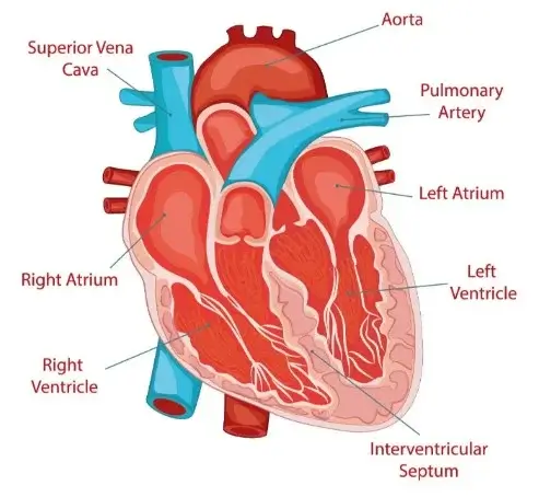
A muscle, the heart pumps blood round the body while at the same time supplying cells with oxygen and food materials as well as removing waste products. It has two atria and two ventricles. These are its elements and functions:
Atria: The uppermost divisions of the human heart are known as atria (atrium singular). They come in pairs, the right and left. It’s main function is to receive blood returning to the heart.
Ventricles: Lower chambers of the heart, which form ventricles. These encompass two types of ventricles including; right ventricle and left ventricle. This passage describes how all organs’ blood circulates through them.
Valves: Valves in one’s heart work normally by redirecting the flow of blood allowing it to move only in one direction thus preventing it from returning to any chamber in his own heart. They look as follows;
- Tricuspid Valve placed between right atrium and right ventricle.
- Pulmonary Valve situated between right ventricle and pulmonary artery.
- Mitral Valve (Bicuspid Valve) found amid left atrium and left ventricle.
- Aortic valve: Found between the left ventricle and the aorta.
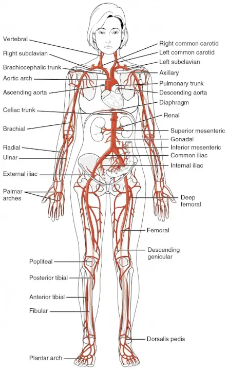
Blood Vessels:
In humans there are blood vessels that help in transportation of fluid mainly through circulatory system. The three types include:
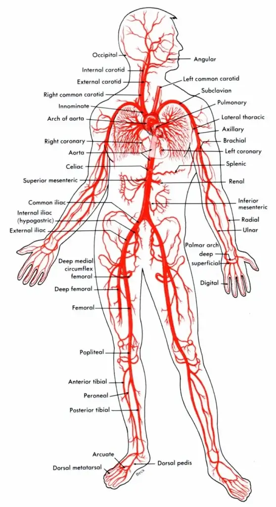
Arteries: They drain away all waste products from tissues and organs back to lungs where they get re-oxygenated. These tubes have very thick walls designed to cope with high pressures caused by pumping action in hearts. Smaller branches of arteries make up arterioles basically.
Veins: Unlike arteries, they are the one which bring back blood with insufficient oxygen to the heart from the organs and tissues of body. Vein walls are thinner compared to arterioles and they have valve that ensures no backflow of blood.
Capillaries: Capillaries are tiny vessels that link arterial systems to venous ones. They assist in gaseous exchange, transport nutrients and remove waste materials between an organism’s cells and its thin-walled circulatory system.
Blood
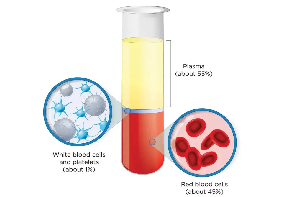
The bodily fluid known as blood is specialized and thus moves via the cardiovascular or circulatory system supplying vital substances including oxygen and essential nutrients to all body cells while at the same time getting rid of unwanted waste materials. It is made up of several parts:
Red Blood Cells (Erythrocytes): These are small doughnuts without holes which form about ninety-nine percent of the population in your bloodstream; their main work it to take life giving air from the lungs over to your body parts like tissues and organs with them containing a protein called hemoglobin that binds with oxygen making our red blood cells red.
White Blood Cells (Leukocytes): White Blood Cells help in fighting off infections due a process called immunity. Different types include macrophages which get rid of pathogens by engulfing them, plasma cells which produce antibodies against invading microbes, neutrophils which coordinate immune response amongst others.
Thrombocytes are blood cells with platelets. It is involved in coagulation (blood clotting). Platelets become adherent to vessels when they get damaged and then secrete factors that initiate clotting, leading to the formation of a clot which arrests further bleeding.
Plasma is about 55% of the volume of blood. It is mostly water and contains various proteins, electrolytes, hormones, nutrients, and waste products. The plasma functions in the transportation of molecules within an organism.
Blood Types
The presence or absence of specific antigens on the surface of red blood cells determines blood types; they are divided into four main groups under the most commonly used classification scheme – ABO system namely; A, B, AB and O. Rh factor on the other hand classifies blood as either positive or negative depending on whether there is presence or absence of the Rh antigen.
Lungs
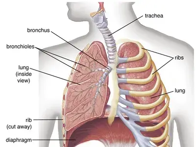
Each of the lungs is a paired organ shaped like a cone and positioned inside the thoracic cavity on every side of the heart. It is enveloped by a two-layered pleura which ensures that it is protected and cushioned.
Bronchi and bronchioles: the trachea splits into two major bronchi (singular: bronchus), one leading to each lung. Further subdivision of these bronchi leads to smaller bronchioles that eventually end up in alveoli and small air sacs.
Alveoli: These are little grape-like sacs where the gas exchange happens. They surround them with capillaries allowing oxygen from air to pass through into blood while carbon dioxide in blood diffuses outside for exhalation via alveoli.
Function
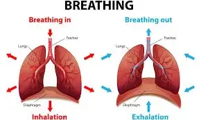
Gas Exchange: The lungs’ main role is oxygen and carbon dioxide between gas and blood vessels. Inhaled oxygen crosses thin walls of alveoli moving toward capillary’s binding with red cells responsible for carrying it to tissues while simultaneously, waste product carbon dioxide from cellular metabolic actions passes out of bloodstream towards alveoli in order to be breathed out.
Ventilation: Ventilation also involves the lungs; a process of breathing. Inhaling, when it is done by us, causes the chest cavity to expand this happens when diaphragm and intercostal muscles contract and air is drawn into the lungs. When these muscles relax, exhalation takes place causing chest cavity to decrease in size expelling air from the lungs at the same time.
Respiratory System Coordination: To regulate breathing rate and maintain proper gas exchange, some other components of respiratory system like diaphragm, rib cage and respiratory centers in brainstem work together with the lungs.
Spleen
The spleen is an important organ that lies in upper left part of abdomen just below your rib cage behind your stomach which belongs to lymphatic system and serve several remarkable functions within human body:
Immune Function: Filtering blood in humans whereby it gets rid of old or damaged red blood cells as well as bacteria, viruses and other pathogens is carried out by spleen. There are specific immune cells called lymphocytes which take part helping the body identify and kill external attackers therefore aiding its immune response.
Blood Storage: The spleen’s role is to store blood for times when its volume may drop due to excessive bleeding or dehydration; it can then release the stored red blood cells and platelets into circulation so as to maintain the necessary level of blood.
Blood Filtration: The function of the spleen regarding blood filtration means that while passing through this organ, substances are removed which include debris, cellular waste products and particles of foreign matter, thus ensuring a good quality of circulating blood and supplying the latter with only healthy blood cells.
Hematopoiesis: In fetal life, the spleen contributes to erythropoiesis. Despite a reduction in its hematopoietic activity at birth, splenic hemopoiesis may still occur under certain conditions such as severe anemia.
Storage of Iron: Also; within this organ, a part of iron reserves in one’s body required for production of red blood cells and several metabolic processes is reserved.
Thymus
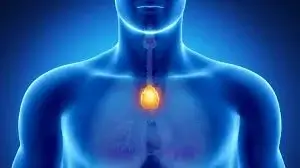
The thymus occupies a special place among primary lymphoid organs located behind sternum in upper chest cavity between lungs. It takes an active part in development and maturation of some immune cells especially T lymphocytes (T cells), which have irreplaceable role in adaptive immunity.
Key features of the thymus include:
Structure: The thymus gland is made up of two lobes, and fibrous tissue separates it into small masses called lobules. A dense outer cortex and a looser inner medulla make up each lobule.
Function:
T Cell Maturation: The thymus gland is the primary lymphoid organ for T cell maturation as well as their selection process to ensure recognition of foreign antigen while avoiding self-reactivity (self-tolerance).
Thymic Education: While T cells mature, they are exposed to peptides associated with MHC molecules presented by specialized TECs called cortical epithelial cells. This exposure helps inform developing T cells about self from non-self antigens thus preventing autoimmunity.
Hormonal Influence: Control on the size of the thymus comes from hormones such as thymosin produced by the thymic epithelium in particular. They contribute to development of T cells.
Involution: In aging individuals’ bodies, the thymus undergoes a process called atrophy. It means that it gradually reduces in size and function, thus leading to less production of new T-cells. Nevertheless, even after maturing into adulthood, maintaining t-cell populations remains to be one potential role that the organ plays.
The circulatory system lacks the presence of bone marrow but it contributes in the formation of blood cells that are essential for functioning of the circulatory system.
Bone Marrow
Bone marrow is not considered a direct part of the circulatory system, but it plays a vital role in the production of blood cells, which are integral components of the circulatory system.
It has two forms:
Red Bone Marrow: Hematopoiesis takes place in red bone marrow to produce all blood cells. The cells known as haematopoietic stem cells (HSCs) residing there differentiate into three basic types of cells namely erythrocytes, leukocytes and platelets. Such blood goes into the bloodstream where it moves about and carries out its functions.
Yellow Bone Marrow: Yellow bone marrow consists mostly of adipocytes; fat-storing cells, which serve as energy stores. Unlike red bone marrow, yellow does not secrete any active blood-forming cells. However, it can reverse back to red marrow in some circumstances e.g., increased requirements for production of blood cells or a disorder affecting this state.
FAQs about circulatory system diagram
What can we learn from the diagram of circulatory system?
Under normal circumstances, a chart showing the basic structure and operation of the heart will provide. The diagram might also indicate other body parts such as the blood vessels (veins, capillaries, arteries) and occasionally the lungs.
How does blood flow through the circulatory system according to the diagram?
Commonly, first the blood is pumped through arteries that are distributed all over in body parts from where it collects in veins returning back to heart. Additionally, it can depict pulmonary circulation indicating that oxygenated blood passes into lungs before going back to heart.
What are some main elements of circulatory systems depicted in diagram?
In general terms these would consist of: pump represented by human heart; arterial system carrying oxygenated blood away from heart; venous one returning deoxygenated blood back to this organ; capillaries facilitating exchange between tissues and plasma.
How does one differentiate among different types of vessels as shown on this drawing?
Arteries are displayed as more massive channels usually filled with bright red fluid moving away from central organ. Veins tend to be portrayed as little tubes transporting liquid stream towards pump.
Explore Related Resources
For a comprehensive overview of how to protect your family in 2026, be sure to read our Ultimate Guide to Digital Parenting.
Additionally, check out these related articles to further secure your household:
- Family Threat Protection Software: Your Digital Shield
- Child Cyber Security Software: The Ultimate Parent’s Guide
- Implantation Cramps: Source of Discomfort, Mean Symptoms, Duration

Russell F. Jones, holding a Master in psychology from the University of Florida. He writes for Smart Parent Solutions, offering practical advice on parenting and child development. His engaging content helps parents navigate family life with confidence and ease. Russell enjoys sharing his knowledge and spending quality time with his family.